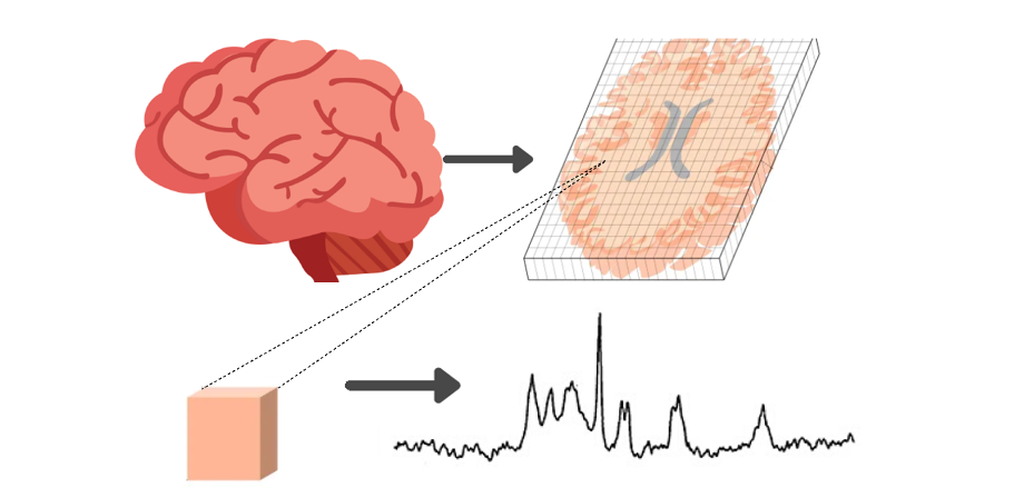
Mapping brain metabolites
The PIPs (Proteins of the Immune system in Psychosis) study will acquire brain imaging scans called magnetic resonance spectroscopy (MRS) that measure the level of small metabolites in brain tissue. Unlike previous MRS studies that measure brain metabolites in single voxels or brain slices based on prior regions, the current study will acquire the brain metabolites at every voxel of the brain. This will allow various brain metabolites, including those indexing neuropil contraction and expansion, oxidative stress and energy metabolism, to be mapped across the entire cortical surface. This is important as the timing and level of brain metabolites varies across brain regions. We are seeking an enthusiastic student to work on advanced methods to quantify the MRS spectra to generate the metabolite maps across the cortex. This work will be done in collaboration with researchers within the Department of Radiology and the University of Geneva. This project is suitable for students with a background in engineering or neuroimaging.